BPG BLOG2

Rare Disease Highlight: Stargardt Disease

Stargardt Disease, also known as juvenile macular degeneration, is a rare disease of the eyes in which part of the eye called the retina does not function correctly. This is the most common form of inherited macular degeneration, affecting about 30,000 people in the United States2. Stargardt’s is a genetic disease caused by a change in the gene ABCA4, which affects how the body uses vitamin A (retinol)4. Normally, vitamin A is helpful to vision because it maintains a clear cornea and is crucial in allowing us to see in low lighting. As a result, fatty materials build up in the part of the eye called the macula, which is a very small part of the retina responsible for seeing sharp images that are straight ahead4. This disease usually starts in childhood but some people do not experience loss of vision until adulthood. Since central vision is compromised, these patients rely on their peripheral vision for most of their sight. Individuals with this disease experience loss of vision, often have trouble going from light to dark places, or have a higher sensitivity to light4. These patients are unable to drive, have trouble reading normal size print, and struggle identifying facial features5. There is no current treatment for Stargardt Disease. Visual aids such as glasses, magnifiers, and wearable visual devices are helpful for these patients, as well as auditory features such as Siri, talk-to-text, and audiobooks1. Even with these aids, patients are not able to see with 20/20 vision, drive a car, or adjust for light sensitivity. This encourages development of new therapies for Stargardt Disease.

References

  1. American Academy of Opthamology. (2022). Resources and Links for People with Low Vision. Retrieved 8/23/22 from https://www.aao.org/eye-health/diseases/low-vision-resources
  2. Foundation Fighting Blindness. (2022). Stargardt Disease. Retrieved 8/19/2022 from https://www.fightingblindness.org/diseases/stargardt-disease#:~:text=Stargardt%20Disease%20is%20the%20most,the%20retina%20called%20the%20macula.
  3. Healthline. (2018). The 9 Most Important Vitamins for Eye Health Retrieved 8/24/22 from https://www.healthline.com/nutrition/eye-vitamins#:~:text=Vitamin%20A,low%20light%20conditions%20(%201%20).
  4. National Eye Institute. (2021). Stargardt Disease. Retrieved 8/19/2022 from https://www.nei.nih.gov/learn-about-eye-health/eye-conditions-and-diseases/stargardt-disease#:~:text=Stargardt%20disease%20is%20a%20rare,vision%20until%20they’re%20adults.
  5. National Library of Medicine. (2010). Stargardt Macular Degeneration. Retrieved 8/23/22 from https://medlineplus.gov/genetics/condition/stargardt-macular-degeneration/

BioPharma Global is a mission-driven corporation dedicated to using our FDA and EMA regulatory expertise and knowledge of various therapeutic areas to help drug developers advance treatments for the disease communities with a unmet medical needs. If you are a drug developer seeking regulatory support for Orphan Drug designation, Fast Track designation, Breakthrough Therapy designation, other FDA/EMA expedited programs, type A, B (pre-IND, EOPs), or C meeting assistance, or IND filings, the BioPharma Global team can help. Contact us today to arrange a 30-minute introductory call.

BPG

Rare Disease Highlight: Netherton Syndrome

Netherton syndrome (NS), also referred to as bamboo hair syndrome, is a rare and severe genetic skin disorder [1-3]. The disease is characterized by chronic ichthyosiform erythroderma (a condition of fine white and red scales across the entire body), atopy (a genetic tendency to develop allergic diseases), and trichorrhexis invaginate (a distinctive hair disorder known as bamboo hair) [1, 2, 4, 5].

NS is a hereditary disorder that arises from a loss-of-function mutation to the serine protease inhibitor Kazal-type 5(SPINK5) gene, which leads to the clinical features of the disease. This type of mutation results in a deficiency of the lympho-epithelial kazal type inhibitor (LEKTI) and increase of specific enzymes known as proteases, such as kallikrein 7 (Klk7) and enzyme elastase 2 (ELA2), and structural components of the outer layer of the skin, such as pro-filgarrin [6]. These molecules are involved in the balance of fatty acids in the skin, natural peeling of the outer layer skin, activation of the inflammatory process, and structure of the skin [7-11]. Thus, the consequential dysregulation results in the breakdown of the skin barrier [2, 6, 12]. NS is transmitted from parents to their children in an autosomal recessive manner. Autosomal refers to genes located on non-sex chromosomes, such as SPINK5. Each chromosome has two working copies of a gene, inherited from each parent. In recessive genetic diseases, both mutated copies of the gene are needed to induce the disease. Therefore, children must receive a faulty gene from each of their parents for NS to become apparent.

Currently, there are about 150 cases of NS reported in the literature, but the true population size is expected to be higher due to difficulties in diagnosis and overlapping of symptoms with other conditions. Since NS is a hereditary disorder generally observed at birth, newborns and infants are the most vulnerable [13]. This patient population fails to thrive in the first year of life. Specifically, newborns with NS manifesting generalized and severe inflammation of the entire skin surface, referred to as congenital ichthyosiform erythroderma (CIE), experience 10-40% mortality in the first year of life. The increased mortality is due to skin barrier disturbances enhancing the risk of infections and hypernatremic dehydration, a form a dehydration where there is too much sodium in the blood [14-16]. Young NS patients also suffer from growth retardation and short stature due to recurrent infections and inflammation [4]. As the patient grows older, the skin disease gradually regresses into a milder form, referred to as ichthyosis linearis circumflexa (ILC). ILC is characterized by recurrent red plaques on the skin with a double scaling edge. Other allergic symptoms caused by NS include asthma, sneezing, congestion, and itchy nose, eczema (a skin condition characterized by dry, itchy, and inflamed skin), and frequent sensitizations to airborne or food allergens [4, 17].

Currently, no satisfactory treatments are approved for NS. Varying success is observed when patients are treated with topical corticosteroids, topical calcineurin inhibitors, topical retinoids, narrowband ultraviolet B phototherapy, ultraviolet radiation, and oral acitretin. For patients with severe disease, intravenous immunoglobulin and anti-tumor necrosis factor-alpha (TNF-α) antibodies may be used [12]. Overall, based on the clinical severity and limited treatments available to effectively treat manifestations of NS, novel therapies for this patient population are needed to address the unmet medical need.

References

1.           OMIM. Netherton Syndrome; NETH. 2012  [cited 2012; Available from: https://www.omim.org/entry/256500.

2.           Leclerc-Mercier, S., et al., Skin Biopsy in Netherton Syndrome: A Histological Review of a Large Series and New Findings. Am J Dermatopathol, 2016. 38(2): p. 83-91.

3.           NIH. Netherton Syndrome. 2019  08/08/2019]; Available from: https://ghr.nlm.nih.gov/condition/netherton-syndrome#statistics.

4.           Hovnanian, A., Netherton syndrome: new advances in the clinic, disease mechanism and treatment AU – Hovnanian, Alain. Expert Review of Dermatology, 2012. 7(1): p. 81-92.

5.           OMIM. Netherton Syndrome. 2019  08/08/2019]; Available from: https://omim.org/entry/256500.

6.           Bonnart, C., et al., Elastase 2 is expressed in human and mouse epidermis and impairs skin barrier function in Netherton syndrome through filaggrin and lipid misprocessing. J Clin Invest, 2010. 120(3): p. 871-82.

7.           Elias, P.M., Y. Hatano, and M.L. Williams, Basis for the barrier abnormality in atopic dermatitis: outside-inside-outside pathogenic mechanisms. J Allergy Clin Immunol, 2008. 121(6): p. 1337-43.

8.           Caubet, C., et al., Degradation of corneodesmosome proteins by two serine proteases of the kallikrein family, SCTE/KLK5/hK5 and SCCE/KLK7/hK7. J Invest Dermatol, 2004. 122(5): p. 1235-44.

9.           Hachem, J.P., et al., Sustained serine proteases activity by prolonged increase in pH leads to degradation of lipid processing enzymes and profound alterations of barrier function and stratum corneum integrity. J Invest Dermatol, 2005. 125(3): p. 510-20.

10.         Nylander-Lundqvist, E., O. Back, and T. Egelrud, IL-1 beta activation in human epidermis. J Immunol, 1996. 157(4): p. 1699-704.

11.         Brattsand, M. and T. Egelrud, Purification and characterization of interleukin 1 beta from human plantar stratum corneum. Evidence of interleukin 1 beta processing in vivo not involving interleukin 1 beta convertase. Cytokine, 1998. 10(7): p. 506-13.

12.         Saleem, H.M.K., et al., Netherton Syndrome: A Case Report and Review of Literature. Cureus, 2018. 10(7): p. e3070-e3070.

13.         NORD. Ichthyosis, Netherton Syndrome. 2005  08/08/2022]; Available from: https://rarediseases.org/rare-diseases/ichthyosis-netherton-syndrome/#:~:text=Netherton%20syndrome%20is%20a%20rare,as%20an%20autosomal%20recessive%20trait.

14.         Stoll, C., et al., Severe hypernatremic dehydration in an infant with Netherton syndrome. Genet Couns, 2001. 12(3): p. 237-43.

15.         Di, W.L., et al., Ex-vivo gene therapy restores LEKTI activity and corrects the architecture of Netherton syndrome-derived skin grafts. Mol Ther, 2011. 19(2): p. 408-16.

16.         Hoeger, P.H. and J.I. Harper, Neonatal erythroderma: differential diagnosis and management of the “red baby”. Archives of Disease in Childhood, 1998. 79(2): p. 186.

17.         Sun, J.D. and K.G. Linden, Netherton syndrome: a case report and review of the literature. Int J Dermatol, 2006. 45(6): p. 693-7.

BioPharma Global is a mission-driven corporation dedicated to using our FDA and EMA regulatory expertise and knowledge of various therapeutic areas to help drug developers advance treatments for the disease communities with a unmet medical needs. If you are a drug developer seeking regulatory support for Orphan Drug designation, Fast Track designation, Breakthrough Therapy designation, other FDA/EMA expedited programs, type A, B (pre-IND, EOPs), or C meeting assistance, or IND filings, the BioPharma Global team can help. Contact us today to arrange a 30-minute introductory call.
BPG BLOG highlight

Rare Disease Highlight: Alternating Hemiplegia of Childhood

Alternating Hemiplegia of Childhood (AHC) is a rare and severe neurodevelopmental disorder that fully manifests during early childhood, typically within the first year of life. AHC is characterized by repeated attacks of hemiplegia, paralysis of one side of the body, or episodes of quadriplegia, simultaneous paralysis of both sides of the body (Heinzen et al., 2014; NORD, 2016; Panagiotakaki et al., 2010; Rosewich et al., 2017; Sweney et al., 2009). The duration of the episodes of paralysis vary, but can last for minutes up to weeks at a time in some cases (NORD, 2016). AHC occurs in approximately 1 in every 1,000,000 births (NORD, 2016).

The symptoms of AHC are diverse and can vary greatly between patients (NORD, 2016). The key hallmark of AHC is the appearance of recurring episodes of paralysis, known as plegic episodes (either hemiplegia or quadriplegia). Other symptoms include tonic attacks (lack of muscle tone), dystonia (painful involuntary muscle contractions), ataxia (lack of coordination), eye problems including nystagmus (uncontrollable eye movements) and strabismus (misalignment of the eyes), developmental delays, neurologic abnormalities, difficulty breathing (dyspnea), and seizures (AHCF, 2022; Panagiotakaki et al., 2010; Sweney et al., 2009). Patients can also develop epilepsy (a brain disorder that causes recurrent and unprovoked seizures) as they get older (NORD, 2016). Symptoms are episodic, range in severity and duration, from minutes to days, and are typically relieved with sleep (AHCF, 2022; Sweney et al., 2009). AHC attacks are triggered by environmental stress, water exposures, specific physical activities, lighting changes, and certain foods (AHCF, 2022; Sweney et al., 2009). During the plegic attacks, patients remain conscious, so the attacks are accompanied by painful vocalization and respiratory compromise (Panagiotakaki et al., 2010; Save et al., 2013; Silver et al., 1993). The prevalence of plegic attacks decreases moderately with age, however these episodic events persist into adulthood (Panagiotakaki et al., 2010).

The cause of AHC is the topic of much research and investigation. In most patients, approximately 76%, AHC is associated with a new sporadic mutation in the ATP1A3 gene (AHCF, 2022; Heinzen et al., 2014; Rosewich et al., 2017). This gene produces the Sodium-Potassium-Transporting ATPase Subunit Alpha 3 protein, required for the normal functioning of neurons (brain nerve cells). Loss of ATPase activity is the root cause of AHC. However, some AHC patients do not present mutations in the ATP1A3 gene, thus scientists believe mutations in other genes could also be associated with AHC onset. Even though AHC appears due to genetic mutations, it is not commonly transmitted from parents to their children. However, in rare cases, AHC can run in families (NORD, 2016).

Patients with AHC experience a reduced quality of life. Patients suffer complications including epilepsy, cognitive impairment, persistent movement disorder, developmental delays, learning disabilities, behavioral issues, sleep disorders, and motor deterioration, all of which appear during childhood and remain into adulthood (Duke Health, 2021; Mikati et al., 2000; Sweney et al., 2009). While life expectancy is not generally affected, children with AHC are at higher risk of life-threatening complications such as breathing difficulties (AHCF, 2022).There are currently no FDA-approved therapies for the treatment of AHC. However, symptoms of AHC can be managed by avoiding triggers and using sleep as a management tactic. AHC attacks and epileptic episodes may also be treated with pharmacologic intervention. The only drug proven effective in reducing the frequency and severity of AHC attacks is the calcium channel entry blocker Flunarizine (approved in Canada). However, it is not effective in all cases and is not readily available in the US (AHCF, 2022; Duke Health, 2021; Kansagra et al., 2013; Neville et al., 2007; NORD, 2016). Therefore, there exists an urgent unmet medical need for patients and caregivers burdened by the profound morbidities that accompany AHC. The development of a therapy administered soon after diagnosis that prevents or reduces the frequency of plegic and dystonic attacks may improve long-term outcomes of patients over the course of their lifetime.

References

AHCF. (2022). Alternating Hemiplegia of Childhood Foundation – What is AHC? Retrieved July 12, 2022 from http://ahckids.org/education/whatisahc/

Duke Health. (2021). Alternating Hemiplegia of Childhood. Retrieved July 12, 2022 from https://www.dukehealth.org/treatments/pediatric-neurology/alternating-hemiplegia-of-childhood

Heinzen, E. L., Arzimanoglou, A., Brashear, A., Clapcote, S. J., Gurrieri, F., Goldstein, D. B., Jóhannesson, S. H., Mikati, M. A., Neville, B., Nicole, S., Ozelius, L. J., Poulsen, H., Schyns, T., Sweadner, K. J., van den Maagdenberg, A., & Vilsen, B. (2014). Distinct neurological disorders with ATP1A3 mutations. Lancet Neurol, 13(5), 503-514. doi:10.1016/s1474-4422(14)70011-0

Kansagra, S., Mikati, M. A., & Vigevano, F. (2013). Alternating hemiplegia of childhood. Handb Clin Neurol, 112, 821-826. doi:10.1016/b978-0-444-52910-7.00001-5

Mikati, M. A., Kramer, U., Zupanc, M. L., & Shanahan, R. J. (2000). Alternating hemiplegia of childhood: clinical manifestations and long-term outcome. Pediatr Neurol, 23(2), 134-141. doi:10.1016/s0887-8994(00)00157-0

Neville, B. G., & Ninan, M. (2007). The treatment and management of alternating hemiplegia of childhood. Dev Med Child Neurol, 49(10), 777-780. doi:10.1111/j.1469-8749.2007.00777.x

NORD. (2016). National Organization for Rare Diseases – Alternating Hemiplegia of Childhood. Retrieved July 12, 2022 from https://rarediseases.org/rare-diseases/alternating-hemiplegia-of-childhood/

Panagiotakaki, E., Gobbi, G., Neville, B., Ebinger, F., Campistol, J., Nevsímalová, S., Laan, L., Casaer, P., Spiel, G., Giannotta, M., Fons, C., Ninan, M., Sange, G., Schyns, T., Vavassori, R., Poncelin, D., & Arzimanoglou, A. (2010). Evidence of a non-progressive course of alternating hemiplegia of childhood: study of a large cohort of children and adults. Brain, 133(Pt 12), 3598-3610. doi:10.1093/brain/awq295

Rosewich, H., Sweney, M. T., DeBrosse, S., Ess, K., Ozelius, L., Andermann, E., Andermann, F., Andrasco, G., Belgrade, A., Brashear, A., Ciccodicola, S., Egan, L., George, A. L., Jr., Lewelt, A., Magelby, J., Merida, M., Newcomb, T., Platt, V., Poncelin, D., Reyna, S., Sasaki, M., Sotero de Menezes, M., Sweadner, K., Viollet, L., Zupanc, M., Silver, K., & Swoboda, K. (2017). Research conference summary from the 2014 International Task Force on ATP1A3-Related Disorders. Neurol Genet, 3(2), e139. doi:10.1212/nxg.0000000000000139

Save, J., Poncelin, D., & Auvin, S. (2013). Caregiver’s burden and psychosocial issues in alternating hemiplegia of childhood. European Journal of Paediatric Neurology, 17(5), 515-521. doi:https://doi.org/10.1016/j.ejpn.2013.04.002

Silver, K., & Andermann, F. (1993). Alternating hemiplegia of childhood: a study of 10 patients and results of flunarizine treatment. Neurology, 43(1), 36-41. doi:10.1212/wnl.43.1_part_1.36

Sweney, M. T., Silver, K., Gerard-Blanluet, M., Pedespan, J.-M., Renault, F., Arzimanoglou, A., Schlesinger-Massart, M., Lewelt, A. J., Reyna, S. P., & Swoboda, K. J. (2009). Alternating Hemiplegia of Childhood: Early Characteristics and Evolution of a Neurodevelopmental Syndrome. Pediatrics, 123(3), e534-e541. doi:10.1542/peds.2008-2027

BioPharma Global is a mission-driven corporation dedicated to using our FDA and EMA regulatory expertise and knowledge of various therapeutic areas to help drug developers advance treatments for the disease communities with a unmet medical needs. If you are a drug developer seeking regulatory support for Orphan Drug designation, Fast Track designation, Breakthrough Therapy designation, other FDA/EMA expedited programs, type A, B (pre-IND, EOPs), or C meeting assistance, or IND filings, the BioPharma Global team can help. Contact us today to arrange a 30-minute introductory call.
BPG BLOG (7)

Rare Disease Highlight: Buruli Ulcer

Buruli ulcer (BU) is a chronic skin infection caused by Mycobacterium ulcerans (M. ulcerans) resulting in death of body tissue, also referred to as necrosis (Scherr et al., 2018). M. ulcerans is the third most common mycobacterial infection worldwide and is classified as a neglected tropical disease by the World Health Organization (WHO) (O’Brien et al., 2018). Cases ofBU have occurred in at least 33 countries, with the highest concentration of cases in Western Africa (Guarner, 2018). M. ulcerans cases are also found in Australia (Guarner, 2018; Thomas et al., 2014). BU is not endemic to the US, however, rare cases have occurred in travelers returning from endemic regions (Thomas et al., 2014). The exact mode of transmission of BU is unknown, and studies have investigated potential risk factors such as insect bites and injuries occurring near bodies of water contaminated with M. ulcerans. BU is not considered a contagious disease, as there are no known cases of human-to-human transmission (Guarner, 2018; Thomas et al., 2014).

M. ulcerans mycobacteria produce a toxin, mycolactone, which diffuses out of the mycobacteria and accumulates in important cells in the body, such as fibroblasts and macrophages (Guarner, 2018). Fibroblasts contribute to the formation of connective tissue that supports and connects other tissues or organs in the body, while macrophages are critical for immunity and tissue development and repair (NIH, 2022; Watanabe et al., 2019). Thus, mycolactone targets and binds to scaffolding and other proteins, disrupting cellular structure and function (Guarner, 2018). The downstream effects of the mycolactone toxin can lead to BU. BU starts as a lesion, which progresses to extensive skin ulceration. Early stages of the disease are significantly painful, but as the disease progresses, the lesional loss of tissue is painless or with limited pain. This is thought to be a result of mycolactone activating type 2 angiotensin II receptors (Yotsu et al., 2018). These receptors are associated with regulating pain, so activation decreases sensations to the skin (Pulakat et al., 2020; Yotsu et al., 2018). Patients left untreated have a risk of disease progression resulting in high morbidity, including physical deformities from skin loss and permanent disability as a result of the spread of necrotizing skin infection to bones and joints (O’Brien et al., 2018).

Effective therapeutic options for BU are limited in the US and worldwide. Patients diagnosed and treated in early stages of the disease have a good prognosis and treatment outcome. However, patients left untreated have a risk of disease progression resulting in high morbidity and permanent disability (O’Brien et al., 2018). Standard of care treatment for BU consists of a combination of antibiotics and complementary treatments. The combination of rifampicin (RIF, 10 mg/kg once daily) and clarithromycin (CLR, 7.5 mg/kg twice daily) is now the recommended treatment by the WHO (WHO, 2022). Despite the success of the RIF+CLR combination therapies, these regimens still require 2 months of treatment, often resulting in patient or provider nonadherence (Phillips et al., 2020). Considering the severe morbidity of the disease and the limited treatment options for patients with BU, novel therapies are necessary to address the unmet medical need for this patient population.

References

Guarner. (2018). Buruli Ulcer: Review of a Neglected Skin Mycobacterial Disease. Journal of Clinical Microbiology, 56(4), e01507-01517. doi:10.1128/jcm.01507-17

NIH. (2022). Fibroblast. Retrieved 07/11/2022 from https://www.genome.gov/genetics-glossary/Fibroblast#:~:text=A%20fibroblast%20is%20a%20type,the%20structural%20framework%20of%20tissues.

O’Brien, Jeanne, Blasdell, Avumegah, & Athan. (2018). The changing epidemiology worldwide of Mycobacterium ulcerans. Epidemiol Infect, 1-8. doi:10.1017/S0950268818002662

Phillips, Robert, Abass, Thompson, Sarfo, Wilson, Sarpong, Gateau, Chauty, Omollo, Ochieng Otieno, Egondi, Ampadu, Agossadou, Marion, Ganlonon, Wansbrough-Jones, Grosset, Macdonald, Treadwell, Saunderson, Paintsil, Lehman, Frimpong, Sarpong, Saizonou, Tiendrebeogo, Ohene, Stienstra, Asiedu, van der Werf, Osei Mireku, Abotsi, Adu Poku, Asamoah-Frimpong, Osei-Wusu, Sarpong, Konadu, Opoku, Forson, Ndogyele, Ofori, Aboagye, Berko, Amofa, Nsiah, Mensah-Bonsu, Ofori Nyarko, Amoako, Koranteng Tannor, Boakye-Appiah, Dzibordzi Loglo, Sarpong-Duah, Agbavor, Ardent, Yamadjako, Adanmado Gersande, Adeye, Kindjinou, Akpolan, Kiki, Sodjinou, Guegnard, Klis, Velding, Omansen, Ofori-Adjei, Eyangoh, Knell, & Faber. (2020). Rifampicin and clarithromycin (extended release) versus rifampicin and streptomycin for limited Buruli ulcer lesions: a randomised, open-label, non-inferiority phase 3 trial. The Lancet, 395(10232), 1259-1267. doi:10.1016/S0140-6736(20)30047-7

Pulakat, & Sumners. (2020). Angiotensin Type 2 Receptors: Painful, or Not? Front Pharmacol, 11, 571994. doi:10.3389/fphar.2020.571994

Scherr, Bieri, Thomas, Chauffour, Kalia, Schneide, Ruf, Lamelas, Manimekalai, Grüber, Ishii, Suzuki, Tanner, Moraski, Miller, Witschel, Jarlier, Pluschke, & Pethe. (2018). Targeting the Mycobacterium ulcerans cytochrome bc1:aa3 for the treatment of Buruli ulcer. Nature Communications, 9(1), 5370. doi:10.1038/s41467-018-07804-8

Thomas, Bailey, Bhatnagar, Ritter, Emery, Jassim, Hornstra, & George. (2014). Mycobacterium ulcerans infection imported from Australia to Missouri, USA, 2012. Emerg Infect Dis, 20(11), 1876-1879. doi:10.3201/eid2011.131534

Watanabe, Alexander, Misharin, & Budinger. (2019). The role of macrophages in the resolution of inflammation. J Clin Invest, 129(7), 2619-2628. doi:10.1172/jci124615

WHO. (2022). Buruli ulcer (Mycobacterium ulcerans infection). Retrieved 07/01/2022 from https://www.who.int/news-room/fact-sheets/detail/buruli-ulcer-(mycobacterium-ulcerans-infection).

Yotsu, Suzuki, Simmonds, Bedimo, Ablordey, Yeboah-Manu, Phillips, & Asiedu. (2018). Buruli Ulcer: a Review of the Current Knowledge. Curr Trop Med Rep, 5(4), 247-256. doi:10.1007/s40475-018-0166-2

BioPharma Global is a mission-driven corporation dedicated to using our FDA and EMA regulatory expertise and knowledge of various therapeutic areas to help drug developers advance treatments for the disease communities with a unmet medical needs. If you are a drug developer seeking regulatory support for Orphan Drug designation, Fast Track designation, Breakthrough Therapy designation, other FDA/EMA expedited programs, type A, B (pre-IND, EOPs), or C meeting assistance, or IND filings, the BioPharma Global team can help. Contact us today to arrange a 30-minute introductory call.

BPG BLOG (64)

Rare Disease Highlight: Congenital disorder of glycosylation type IIc (CDGIIc)

Leukocyte Adhesion Deficiencies (LAD) are autosomal recessive immunodeficiencies characterized by severe and recurrent bacterial infections, impaired wound healing, and neutrophilia (Cagdas et al., 2014). Congenital disorder of glycosylation type IIc (CDGIIc) is an ultra-rare LAD in which patients exhibit these general LAD phenotypes, but are sometimes further characterized by two additional potential symptoms including moderate to severe psychomotor retardation and mild dysmorphism (OMIM, 2014). In CDGIIc patients, the body is unable to sufficiently add fucose sugars to the surface of certain cells (hypo-fucosylation), which causes leukocytosis, persistent and recurrent infections, severely reduced selectin activity, Bombay blood group, hypotonia, poor feeding, short stature, coarse facial features and neurological deficits. This is mainly because the leukocytes in the body are unable to adhere to cell surfaces which would normally be marked with fucose sugars to fight off infections (Marquardt et al., 1999). In some cases, patients died because of clinical manifestations of the disease (confirmed after death), demonstrating the severe and life-threatening nature of CDGIIc (Cagdas et al., 2014; Karsan et al., 1998).

Immune defects are the direct result of a lack of cluster of differentiation (CD) 15, a fucose-containing, cell surface glycoprotein that serves as a ligand for certain cell adhesion molecules called E- and P‑selectins (Frydman et al., 1996). In addition, CDGIIc patients’ red blood cells lack the H antigen, a fucosylated glycoprotein, which is the precursor molecule of the A, B, and O blood groups, and consequently manifests as the Bombay blood type. Recurrent bacterial infections occur in almost all patients during the first years of life, although periodontitis has been reported at later ages (A. Etzioni et al., 1992). Biochemical analysis confirms global hypo-fucosylation of proteoglycans, suggesting an inborn error of fucose metabolism. CDGIIc is a serious and potentially life-threatening disease, particularly for infants and young children (Dauber et al., 2014; A. Etzioni et al., 1992; Amos Etzioni et al., 2002; Frydman et al., 1992; Karsan et al., 1998; Marquardt et al., 1999; Sturla et al., 1998). The onset of clinical symptoms is typically during infancy and almost always within the first year of life. Without medical intervention, clinical manifestations of CDGIIc, in most cases, persist through development or result in patient mortality during early childhood (Dauber et al., 2014; A. Etzioni et al., 1992; Amos Etzioni et al., 2002; Frydman et al., 1992; Marquardt et al., 1999; Sturla et al., 1998)

References

Cagdas, D., Yilmaz, M., Kandemir, N., Tezcan, I., Etzioni, A., & Sanal, Ö. (2014). A novel mutation in leukocyte adhesion deficiency type II/CDGIIc. Journal of clinical immunology, 34(8), 1009–1014. doi:10.1007/s10875-014-0091-7

Dauber, A., Ercan, A., Lee, J., James, P., Jacobs, P. P., Ashline, D. J., . . . Sackstein, R. (2014). Congenital disorder of fucosylation type 2c (LADII) presenting with short stature and developmental delay with minimal adhesion defect. Human molecular genetics, 23(11), 2880–2887. doi:10.1093/hmg/ddu001

Etzioni, A., Frydman, M., Pollack, S., Avidor, I., Phillips, M. L., Paulson, J. C., & Gershoni-Baruch, R. (1992). Brief report: Recurrent severe infections caused by a novel leukocyte adhesion deficiency. N Engl.J Med, 327(25), 1789–1792. doi:10.1056/nejm199212173272505

Etzioni, A., Sturla, L., Antonellis, A., Green, E. D., Gershoni-Baruch, R., Berninsone, P. M., . . . Tonetti, M. (2002). Leukocyte adhesion deficiency (LAD) type II/carbohydrate deficient glycoprotein (CDG) IIc founder effect and genotype/phenotype correlation. Am J Med Genet, 110(2), 131–135. doi:10.1002/ajmg.10423

Frydman, M., Etzioni, A., Eidlitz-Markus, T., Avidor, I., Varsano, I., Shechter, Y., . . . Gershoni-Baruch, R. (1992). Rambam-Hasharon syndrome of psychomotor retardation, short stature, defective neutrophil motility, and Bombay phenotype. Am J Med Genet, 44(3), 297–302. doi:10.1002/ajmg.1320440307

Frydman, M., Vardimon, D., Shalev, E., & Orlin, J. B. (1996). Prenatal diagnosis of Rambam-Hasharon syndrome. Prenat Diagn, 16(3), 266-269. doi:10.1002/(sici)1097-0223(199603)16:3<266::Aid-pd845>3.0.Co;2-#

Karsan, A., Cornejo, C. J., Winn, R. K., Schwartz, B. R., Way, W., Lannir, N., . . . Harlan, J. M. (1998). Leukocyte Adhesion Deficiency Type II is a generalized defect of de novo GDP-fucose biosynthesis. Endothelial cell fucosylation is not required for neutrophil rolling on human nonlymphoid endothelium. J Clin Invest, 101(11), 2438–2445. doi:10.1172/jci905

Marquardt, T., Brune, T., Lühn, K., Zimmer, K. P., Körner, C., Fabritz, L., . . . Koch, H. G. (1999). Leukocyte adhesion deficiency II syndrome, a generalized defect in fucose metabolism. J Pediatr, 134(6), 681–688.

OMIM. (2014). Online Mendelian Inheritance in Man: MIM Number: 266265. Retrieved from https://omim.org

Sturla, L., Etzioni, A., Bisso, A., Zanardi, D., Flora, G. d., Silengo, L., . . . Tonetti, M. (1998). Defective intracellular activity of GDP-D-mannose-4,6-dehydratase in leukocyte adhesion deficiency type II syndrome. FEBS Lett, 429(3), 274–278.

BioPharma Global is a mission-driven corporation dedicated to using our FDA and EMA regulatory expertise and knowledge of various therapeutic areas to help drug developers advance treatments for the disease communities with a unmet medical needs. If you are a drug developer seeking regulatory support for Orphan Drug designation, Fast Track designation, Breakthrough Therapy designation, other FDA/EMA expedited programs, type A, B (pre-IND, EOPs), or C meeting assistance, or IND filings, the BioPharma Global team can help. Contact us today to arrange a 30-minute introductory call.
BPG BLOG (3)

Rare Disease Highlight: Acinetobacter infection

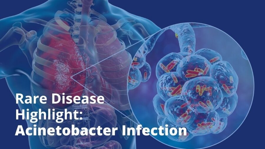
Acinetobacter infection is a rare and serious bacterial disease acquired by combat troops returning from conflict zones in areas of the Middle East (Howard et al., 2012). In addition, Acinetobacter infection occurs in intensive care unit (ICU) patients who are critically ill or have a weakened immune system (CDC, 2010). The disease can initially manifest as nosocomial pneumonia, bloodstream or wound infection, urinary tract infection, or other type of opportunistic infection. Across the range of anatomical regions which may be involved, Acinetobacter infections are typically associated with worse outcomes compared with more commonly isolated organisms. For example, ventilator associated pneumonia (VAP) is defined as occurring for more than 48 hours following mechanical ventilation in an intubated patient, and accounts for 86% of all nosocomial pneumonias. VAP caused by Acinetobacter is associated with mortality rates up to 70% and greater disease severity than other etiologies (Howard et al., 2012; Koenig et al., 2006). Acinetobacter infections also account for 1.3% of all nosocomial bloodstream infections in the US and are associated with the third highest mortality rate in the ICU below Pseudomonas infection and Candidiasis.

Normally found in aquatic environments, Acinetobacter can be found in cultures of hospitalized patients’ sputum, respiratory secretions, wounds, and urine, but normally has low virulence (Cunha, 2016; Howard et al., 2012). The genus Acinetobacter includes a heterogeneous group of 34 formally named species of aerobic nonhemolytic Gram-negative coccobacilli, which are usually found in diploid formation or chains of variable length. Among the Acinetobacter species, A. baumanniiA. pittii, and A. nosocomialis are closely related and are considered important nosocomial pathogens accounting for most Acinetobacter infections (Kim et al., 2014). Of these, Acinetobacter baumannii accounts for 80% of reported infections (CDC, 2010).

A. baumannii is a Gram-negative, pleomorphic, aerobic, catalase-positive, oxidase-negative, non-motile, non-fermenting coccobacillus. Carbapenem-resistant Acinetobacter baumannii (CRAB) refers to strains of this pathogen which have reduced susceptibility to carbapenems due to a variety of mechanisms including expression of a carbapenemase enzyme, porin loss and/or changes in membrane permeability (Codjoe et al., 2018). This activity contributes to the high frequency of multidrug-resistance (MDR) and the high severity of infections caused by CRAB (Queenan et al., 2007). The incidence of MDR properties among all A. baumannii infections is estimated from 30-80% of reported cases (CDC, 2011; Xiao et al., 2017).

References

CDC. (2010). Acinetobacter in Healthcare Settings. Retrieved from https://www.cdc.gov/hai/organisms/acinetobacter.html

CDC. (2011). Gram-negative Bacteria Infections in Healthcare Settings. Retrieved from https://www.cdc.gov/hai/organisms/gram-negative-bacteria.html

Codjoe, F. S., & Donkor, E. S. (2018). Carbapenem Resistance: A Review. Medical Sciences, 6(1), 1.

Cunha, B. (2016). Acinetobacter. Retrieved from https://emedicine.medscape.com/article/236891

Howard, A., O’Donoghue, M., Feeney, A., & Sleator, R. D. (2012). Acinetobacter baumannii: an emerging opportunistic pathogen. Virulence, 3(3), 243-250. doi:10.4161/viru.19700

Kim, U. J., Kim, H. K., An, J. H., Cho, S. K., Park, K.-H., & Jang, H.-C. (2014). Update on the Epidemiology, Treatment, and Outcomes of Carbapenem-resistant Acinetobacter infections. Chonnam medical journal, 50(2), 37-44. doi:10.4068/cmj.2014.50.2.37

Koenig, S. M., & Truwit, J. D. (2006). Ventilator-associated pneumonia: diagnosis, treatment, and prevention. Clinical microbiology reviews, 19(4), 637-657. doi:10.1128/CMR.00051-05

Queenan, A. M., & Bush, K. (2007). Carbapenemases: the versatile beta-lactamases. Clinical microbiology reviews, 20(3), 440-458. doi:10.1128/CMR.00001-07

Xiao, D., Wang, L., Zhang, D., Xiang, D., Liu, Q., & Xing, X. (2017). Prognosis of patients with Acinetobacter baumannii infection in the intensive care unit: A retrospective analysis. Experimental and therapeutic medicine, 13(4), 1630-1633. doi:10.3892/etm.2017.4137

BioPharma Global is a mission-driven corporation dedicated to using our FDA and EMA regulatory expertise and knowledge of various therapeutic areas to help drug developers advance treatments for the disease communities with a unmet medical needs. If you are a drug developer seeking regulatory support for Orphan Drug designation, Fast Track designation, Breakthrough Therapy designation, other FDA/EMA expedited programs, type A, B (pre-IND, EOPs), or C meeting assistance, or IND filings, the BioPharma Global team can help. Contact us today to arrange a 30-minute introductory call.
BPG BLOG (52)

Rare Disease Highlight: Fragile X Syndrome

Fragile X Syndrome (FXS) is a dominant genetic disorder that causes a range of symptoms from intellectual disability, developmental delays, and motor dysfunction, to abnormalities in the testes and ovaries (known as gonadal abnormalities) (Garber et al., 2008). While FXS is a rare disease affecting approximately 54,000 people in the US, it is the most common cause of inherited intellectual disability, second in prevalence only to Down syndrome (Coffee et al., 2009; Crawford et al., 1999; Saldarriaga et al., 2014). The cause of FXS is an abnormal expansion of a trinucleotide repeat in the Fragile X Mental Retardation 1 gene (FMR1). Nucleotides are the basic units that compose deoxyribonucleic acid (DNA). A trinucleotide repeat refers to the presence of three nucleotides consecutively repeated within a specific region of the DNA. Typically, the trinucleotide of cytosine-guanine-guanine (CGG) in the FMR1 gene is composed of less than 45 repeats. However, in FXS, the CGG trinucleotide repeats more than 200 times, which is known as a full mutation of FMR1 (Garber et al., 2008). This mutation impairs FMR1’s ability to produce the Fragile X Mental Retardation Protein (FMRP), essential for normal brain development (D’Incal et al., 2022).

The symptoms of FXS vary depending on several characteristics, including how many trinucleotide repeats are present, sex, and magnitude of FMRP deficit (Hagerman et al., 2009; McConkie-Rosell et al., 2005; Merenstein et al., 1996). The FMR1 gene is located on the sex chromosome known as X chromosome. Biologically, females have two X chromosomes while males have only one. Therefore, FXS is more common in males while females present milder symptoms (Verdura et al., 2021). The main symptoms of FXS are (Ciaccio et al., 2017; Lachiewicz et al., 2000; McConkie-Rosell et al., 2005):

  • Intellectual disability
  • Facial deformities, including long narrow face, large head (macrocephaly), and prominent ears and/or jaw
  • Psychomotor dysfunction, including delay in crawling and walking, low muscle tone, poor coordination and balance, spine deformity, and flat feet
  • Social dysfunction, including increased aggressiveness, anxiety, and depression, autism, and sleeping difficulties
  • Organization problems (attention deficit hyperactivity disorder [ADHD])
  • Functional deficits, including lack of hygiene and grooming, and inability to perform household tasks and maintain weight
  • Seizures
  • Heart disease
  • Misalignment of the eyes (strabismus)
  • Recurrent inflammation in the middle ear (otitis media)
  • Gastrointestinal dysfunction

The lifespan of FXS patients is generally unaffected by the disease. However, patients live with significant morbidities such as intellectual disability, functional deficits, social dysfunction, neuropsychological problems, and physical disabilities (Bailey et al., 2009). These morbidities significantly decrease FXS patients’ quality of life since they limit patients’ independence and ability to function without assistance. FXS patients with significant intellectual disability have difficulty learning new tasks, feeding themselves, keeping an organized and/or clean environment, bathing, and dressing. They also have challenges maintaining a satisfactory social life due to their behavioral and social abnormalities. Furthermore, FXS patients display motor dysfunction, restricting daily movement. As a result of the intellectual, social, behavioral, and motor challenges, FXS patients typically require a full-time caregiver and are dependent on others for survival (Ciaccio et al., 2017; Lachiewicz et al., 2000; McConkie-Rosell et al., 2005). Despite the serious nature of FXS, there are no approved curative therapies to date. Treatment of FXS patients focus on symptom management and includes non-pharmacological therapies such as speech and language, behavioral, and physical therapy, as well as pharmacological therapies. The most common drugs used for these patients are stimulants to improve attention deficit and hyperactivity, serotonin reuptake inhibitors to relieve anxiety, and antipsychotics to reduce irritability and aggression (Ciaccio et al., 2017).

FXS has a profound impact in patients’ life and the serious nature of the disease and the lack of effective treatments highlight the unmet medical need for this patient population. Thus, the development of novel pharmacological agents that can improve the quality of life of FXS patients is essential.

References

Bailey, D. B., Jr., Raspa, M., Bishop, E., & Holiday, D. (2009). No change in the age of diagnosis for fragile x syndrome: findings from a national parent survey. Pediatrics, 124(2), 527-533. doi:10.1542/peds.2008-2992

Ciaccio, C., Fontana, L., Milani, D., Tabano, S., Miozzo, M., & Esposito, S. (2017). Fragile X syndrome: a review of clinical and molecular diagnoses. Italian Journal of Pediatrics, 43(1), 39. doi:10.1186/s13052-017-0355-y

Coffee, B., Keith, K., Albizua, I., Malone, T., Mowrey, J., Sherman, S. L., & Warren, S. T. (2009). Incidence of fragile X syndrome by newborn screening for methylated FMR1 DNA. Am J Hum Genet, 85(4), 503-514. doi:10.1016/j.ajhg.2009.09.007

Crawford, D. C., Meadows, K. L., Newman, J. L., Taft, L. F., Pettay, D. L., Gold, L. B., Hersey, S. J., Hinkle, E. F., Stanfield, M. L., Holmgreen, P., Yeargin-Allsopp, M., Boyle, C., & Sherman, S. L. (1999). Prevalence and phenotype consequence of FRAXA and FRAXE alleles in a large, ethnically diverse, special education-needs population. Am J Hum Genet, 64(2), 495-507. doi:10.1086/302260

D’Incal, C., Broos, J., Torfs, T., Kooy, R. F., & Vanden Berghe, W. (2022). Towards Kinase Inhibitor Therapies for Fragile X Syndrome: Tweaking Twists in the Autism Spectrum Kinase Signaling Network. Cells, 11(8). doi:10.3390/cells11081325

Garber, K. B., Visootsak, J., & Warren, S. T. (2008). Fragile X syndrome. European Journal of Human Genetics, 16(6), 666-672. doi:10.1038/ejhg.2008.61

Hagerman, R. J., Berry-Kravis, E., Kaufmann, W. E., Ono, M. Y., Tartaglia, N., Lachiewicz, A., Kronk, R., Delahunty, C., Hessl, D., Visootsak, J., Picker, J., Gane, L., & Tranfaglia, M. (2009). Advances in the treatment of fragile X syndrome. Pediatrics, 123(1), 378-390. doi:10.1542/peds.2008-0317

Lachiewicz, A. M., Dawson, D. V., & Spiridigliozzi, G. A. (2000). Physical characteristics of young boys with fragile X syndrome: reasons for difficulties in making a diagnosis in young males. Am J Med Genet, 92(4), 229-236. doi:10.1002/(sici)1096-8628(20000605)92:4<229::aid-ajmg1>3.0.co;2-k

McConkie-Rosell, A., Finucane, B., Cronister, A., Abrams, L., Bennett, R. L., & Pettersen, B. J. (2005). Genetic counseling for fragile x syndrome: updated recommendations of the national society of genetic counselors. J Genet Couns, 14(4), 249-270. doi:10.1007/s10897-005-4802-x

Merenstein, S. A., Sobesky, W. E., Taylor, A. K., Riddle, J. E., Tran, H. X., & Hagerman, R. J. (1996). Molecular-clinical correlations in males with an expanded FMR1 mutation. Am J Med Genet, 64(2), 388-394. doi:10.1002/(sici)1096-8628(19960809)64:2<388::Aid-ajmg31>3.0.Co;2-9

Saldarriaga, W., Tassone, F., González-Teshima, L. Y., Forero-Forero, J. V., Ayala-Zapata, S., & Hagerman, R. (2014). Fragile X syndrome. Colomb Med (Cali), 45(4), 190-198.

Verdura, E., Pérez-Cano, L., Sabido-Vera, R., Guney, E., Hyvelin, J.-M., Durham, L., & Gomez-Mancilla, B. (2021). Heterogeneity in Fragile X Syndrome Highlights the Need for Precision Medicine-Based Treatments. Frontiers in Psychiatry, 12. doi:10.3389/fpsyt.2021.722378

BioPharma Global is a mission-driven corporation dedicated to using our FDA and EMA regulatory expertise and knowledge of various therapeutic areas to help drug developers advance treatments for the disease communities with a unmet medical needs. If you are a drug developer seeking regulatory support for Orphan Drug designation, Fast Track designation, Breakthrough Therapy designation, other FDA/EMA expedited programs, type A, B (pre-IND, EOPs), or C meeting assistance, or IND filings, the BioPharma Global team can help. Contact us today to arrange a 30-minute introductory call.

BPG BLOG (47)

Rare Disease Highlight: Acute Radiation Syndrome (ARS)

Acute Radiation Syndrome (ARS) is a severe acute illness caused by irradiation of the majority of the body by a high dose of ionizing radiation within a brief timeframe (CDC, 2017). According to the National Council on Radiation Protection and Measurements (NCRP), ARS is defined as “[…]a broad term used to describe a range of signs and symptoms that reflect severe damage to specific organ systems and that can lead to death within hours or up to several months after exposure.” (Acosta et al., 2017). The disease is characterized by depletion of immature stem cells in specific functional tissues exposed to ionizing radiation (CDC, 2017). Hazardous sources of ionizing radiation can include faults in medical imaging technology, a nuclear plant meltdown, or an attack using a nuclear weapon. People within the vicinity of these events become exposed to large amounts of radiation, causing ARS (Acosta et al., 2017).

The severity of ARS is directly linked to radiation dose, with higher doses leading to damage of more sensitive organ systems. Upon contact with intracellular targets or other molecules in the body, ionizing radiation produces free radicals which themselves become highly damaging on interaction with other molecules or tissues. The cells most critically affected by radiation are those which rapidly develop and divide, including hematopoietic stem cells (HSC) in the bone marrow, spermatocytes in the testes, and crypt cells in the intestines (López et al., 2011).

There are several factors which determine the lethality of ionizing radiation, including: dose rate, distance from the radiation source, and shielding. Receiving the same dose of radiation concentrated over a shorter period causes more damage to tissues, but the overall dose rate decreases as the distance from the source increases. Shielding can reduce exposure, depending on the shielding material (López et al., 2011).

As ARS affects multiple organ systems in the body, current treatment and standards of care (SoC) for ARS depend on the affected tissues. The most immediate goals of patient management involve physical exam, removal of external contamination, radiation dose estimation, supportive care (including psychological support of the patient and family), symptomatic treatment, blood transfusions as needed, and replacement of fluids and electrolytes (CDC, 2017). Treatment with certain cell signaling factors known as cytokines such as granulocyte colony stimulating factors (G-CSF) and keratinocyte growth factor may be used to treat other manifestations of radiation exposure (Choi et al., 2017). When required, surgical intervention must be carried out within the first 36-48 hours from exposure, since immunosuppression caused by the radiation increases risk for infection (López et al., 2011). Although ARS is extremely rare in the United States, historical cases of mass radiation exposure serve as an important reminder to prepare for the possibility for ARS in the future.

References

Acosta, R., & Warrington, S. J. (2017). Radiation, Syndrome Acute. In StatPearls. Treasure Island (FL): StatPearls Publishing

StatPearls Publishing LLC.

CDC. (2017). Acute Radiation Syndrome: A Fact Sheet For Clinicians.  Retrieved February 23, 2018 https://emergency.cdc.gov/radiation/arsphysicianfactsheet.asp

Choi, J.-S., Shin, H.-S., An, H.-Y., Kim, Y.-M., & Lim, J.-Y. (2017). Radioprotective effects of Keratinocyte Growth Factor-1 against irradiation-induced salivary gland hypofunction. Oncotarget, 8(8), 13496-13508. doi:10.18632/oncotarget.14583

López, M., & Martín, M. (2011). Medical management of the acute radiation syndrome. Reports of Practical Oncology and Radiotherapy, 16(4), 138-146. doi:10.1016/j.rpor.2011.05.001

RCR. (2016). Radiotherapy dose fractionation, second edition.

BioPharma Global is a mission-driven corporation dedicated to using our FDA and EMA regulatory expertise and knowledge of various therapeutic areas to help drug developers advance treatments for the disease communities with a unmet medical needs. If you are a drug developer seeking regulatory support for Orphan Drug designation, Fast Track designation, Breakthrough Therapy designation, other FDA/EMA expedited programs, type A, B (pre-IND, EOPs), or C meeting assistance, or IND filings, the BioPharma Global team can help. Contact us today to arrange a 30-minute introductory call.

BPG BLOG (42)

Rare Disease Highlight: Spinocerebellar Ataxia

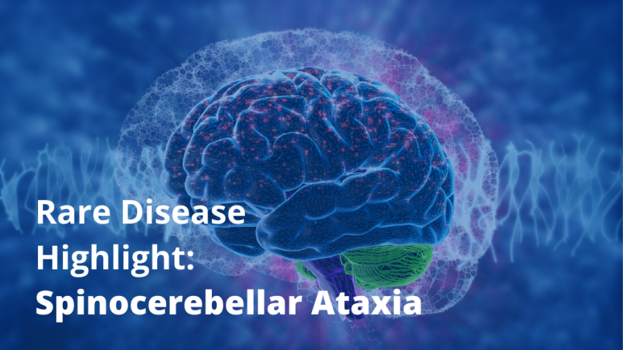
Spinocerebellar ataxia (SCA) is a group of autosomal dominant, progressive neurodegenerative disorders, mainly affecting the cerebellum and/or brain stem (Sullivan et al., 2019).Ataxia is also known as impaired coordination due to damage to the brain, nerves, or muscles. Since SCA is inherited in an autosomal dominant pattern, one parent can pass the mutation to the child, with symptoms observable as early as birth (NAF, 2015). Generally, patients with SCA present with 3 core features (Sullivan et al., 2019):

  1. Ataxic gait – difficulty walking in a straight line, incoordination, veering side-to-side, widened stance for better base support, and inconsistent arm motion.
  2.  Nystagmus/visual problems – involuntary rhythmic eye motions.
  3. Dysarthria – weakened muscles causing slurred speech.

Specific subtypes of SCA can also manifest other symptoms in the trunk and/or limbs (Sullivan et al., 2019):

  1. Extrapyramidal and pyramidal signs – involuntary movements (such as tremors and muscle contractions) and abnormal reactions to external stimuli upon neurological examination, respectively.
  2. Ophthalmoplegia – paralysis of muscles controlling eye movements.
  3. Cognitive impairments, including intellectual disability or dementia.

In the most common subtypes of SCA, a CAG (cytosine, adenine, and guanine) trinucleotide repeat in a specific gene causes an expansion in certain proteins (Pilotto et al., 2018). Nucleotides are the basic units composing deoxyribonucleic acid (DNA). Therefore, a trinucleotide repeat refers to the presence of three nucleotides consecutively repeated within a specific region of the DNA. As a result, these proteins misfold and accumulate in neuronal cells within the cerebellum or spinal cord. The affected neurons progressively become dysfunctional and begin to degrade, manifesting as ataxia (Pilotto et al., 2018). The cause of other SCA subtypes are based on specific genetic variations (Sullivan et al., 2019).

SCA is a physically debilitating disease, leaving patients unable to execute activities of daily living without assistance. As the disease progresses in severity, patients can lose neurological function, potentially leading to intellectual disability, slurred speech, or death from complete brainstem failure. Survival can range from 8 years after symptom onset to a normal lifespan (O’Sullivan et al., 2004). In the most common SCA subtypes, patients are expected to survive for a few decades following initial onset of symptoms (O’Sullivan et al., 2004; Tezenas du Montcel et al., 2014). However, severe cases of subtypes SCA2, SCA3, SCA7, SCA16, and SCA17 have the shortest survival (O’Sullivan et al., 2004). Overall patient prognosis is determined by the length of CAG repeat expansion (Pilotto et al., 2018).

Currently, there is no cure for SCA, so supportive care for symptomatic relief is the goal of treatment. To manage ataxia, patients participate in physical therapy to preserve muscle strength and tone. In addition, patients might receive special mobility-assisting devices, such as a walkers, canes, or wheelchairs. For symptoms such as tremors, muscle stiffness, muscle spasms, and sleep disorders, medications are prescribed as needed (GARD, 2022). SCA patients are recommended to have consistent, long term follow-up with their healthcare professionals to monitor and prevent complications in the heart, lungs, spine, bones, and/or muscles (NORD et al., 2022). Thus, despite available standard of care for symptom management, an unmet medical need for a novel therapy exists for SCA patients.

References

GARD. (2022, 2017). Spinocerebellar Ataxia. Retrieved 4/25/2022 from https://rarediseases.info.nih.gov/diseases/10748/spinocerebellar-ataxia.

NAF. (2015). Classification of Ataxia. National Ataxia Foundation. Retrieved from https://www.ataxia.org/wp-content/uploads/2019/04/Ataxia-Classification.pdf

NORD, & Bird. (2022, 2017). Autosomal Dominant Hereditary Ataxia. Rare Disease Database. Retrieved 4/25/2022 from https://rarediseases.org/rare-diseases/autosomal-dominant-hereditary-ataxia/.

O’Sullivan, Michelson, Bennett, & Bird. (2004). Spinocerebellar Ataxia: Making an Informed Choice About Genetic Testing. Medical Genetics and Neurology. Retrieved from https://www.ataxia.org/wp-content/uploads/2017/07/SCA-Making_an_Informed_Choice_About_Genetic_Testing.pdf

Pilotto, & Saxena. (2018). Epidemiology of inherited cerebellar ataxias and challenges in clinical research. Clinical and Translational Neuroscience, 2(2), 2514183X18785258. doi:10.1177/2514183×18785258

Sullivan, Yau, O’Connor, & Houlden. (2019). Spinocerebellar ataxia: an update. Journal of neurology, 266(2), 533-544. doi:10.1007/s00415-018-9076-4

Tezenas du Montcel, Durr, Rakowicz, Nanetti, Charles, Sulek, Mariotti, Rola, Schols, Bauer, Dufaure-Garé, Jacobi, Forlani, Schmitz-Hübsch, Filla, Timmann, van de Warrenburg, Marelli, Kang, Giunti, Cook, Baliko, Bela, Boesch, Szymanski, Berciano, Infante, Buerk, Masciullo, Di Fabio, Depondt, Ratka, Stevanin, Klockgether, Brice, & Golmard. (2014). Prediction of the age at onset in spinocerebellar ataxia type 1, 2, 3 and 6. Journal of Medical Genetics, 51(7), 479-486. doi:10.1136/jmedgenet-2013-102200

BioPharma Global is a mission-driven corporation dedicated to using our FDA and EMA regulatory expertise and knowledge of various therapeutic areas to help drug developers advance treatments for the disease communities with a unmet medical needs. If you are a drug developer seeking regulatory support for Orphan Drug designation, Fast Track designation, Breakthrough Therapy designation, other FDA/EMA expedited programs, type A, B (pre-IND, EOPs), or C meeting assistance, or IND filings, the BioPharma Global team can help. Contact us today to arrange a 30-minute introductory call.
BPG BLOG (37)

Rare Disease Highlight: Gastric Cancer

Gastric Cancer (GC) is a disease in which malignant cells form into tumors in the lining of the stomach (NCI, 2021). About 90-95% of GC are adenocarcinomas, which develop from the gland cells in the mucosa (innermost lining of the stomach) (ACS, 2021a). With treatment, the 5-year survival rate of early GC can reach up to 95% (Song et al., 2017). However, 5-year survival rate of all stages combined (early to advanced disease) is 32% because there is no standard or routine screening test for GC in the US (ACS, 2021a; NCI, 2021; SEER, 2021). Most patients, ~73%, are diagnosed at an advanced stage with distant GC (AJCC stage IV). These patients have a 5-year survival rate of only 6%. GC is a rare disease that affects approximately 133,000 people in the US (SEER, 2021).

GC is a multifactorial disease because its occurrence and development are influenced by both environmental and genetic factors. Risk factors associated with GC include family history, diet, alcohol consumption, tobacco smoking, and Helicobacter pylori (H. pylori) and Epstein-Barr virus (EBV) infections. Although GC is largely sporadic, family history of GC remains one of the most crucial risk factors, with about 10% of cases displaying familial aggregation. Further, individuals with this family history have 3-fold higher risk of getting gastric carcinoma than individuals without such history (Machlowska et al., 2020). Salt-preserved foods may impact GC progression by inducing changes in the gastric mucosa towards precancerous conditions. Tobacco smoking also plays a role, as smoking is associated with up to 18% of GC cases (Marqués-Lespier et al., 2016). Additionally, there is a positive interaction between smoking and H. pylori infection (Piazuelo et al., 2013). H. pylori affects GC development by two mechanisms: an indirect inflammatory reaction to H. pylori infection on the gastric mucosa and a direct epigenetic outcome of H. pylori on gastric epithelial cells (Machlowska et al., 2020). Lastly, EBV is associated with GC development, although limited evidence exists of EBV-infected white blood cells entering the gastric epithelium (Figueiredo et al., 2017).

The symptoms of GC depend on the stage of the disease. In early stages, patients may experience indigestion, stomach discomfort, a bloated feeling after eating, mild nausea, loss of appetite, and/or heartburn. In advanced stages, patients may experience blood in the stool, vomiting, unexplained weight loss, stomach pain, yellowing of the skin (jaundice), build-up of fluid in the abdomen (ascites), and/or trouble swallowing (NCI, 2021). GC is staged based on the location and extent of the main tumor and classified as Stage 0, I, II, III, or IV, from early disease to advanced disease with metastasis (when the cancer spreads to other parts of the body) (ACS, 2021b; NCI, 2021).

The main treatments for GC are surgery, chemotherapy, targeted drug therapy, immunotherapy, and radiation therapy. Generally, combinations of two or more drugs are used to treat GC, depending on cancer stage, patient’s overall health, and the ability to undergo radiotherapy. Surgery, such as gastrectomy (removal of all or part of the stomach), is a common treatment for all stages of GC. It is used as a curative treatment for stages I-III patients and as a palliative treatment for stage IV patients, as well as patients who relapse (NCI, 2021). Although surgical removal of the stomach offers the potential to cure GC and prolongs patient survival, it significantly affects patients’ quality of life. Following surgery, some patients still need chemotherapy and/or radiation therapy. These treatments can exacerbate nutritional deficiencies. To help with this problem, a jejunostomy tube (a soft, plastic tube placed through the skin of the abdomen into the midsection of the small intestine to deliver food and medicine) is placed into the intestine at the time of or after the surgery to provide liquid nutrition to patients (NCI 2021 b and NIC 2021) (ACS, 2021a). Targeted therapies such as monoclonal antibodies can be used to target specific cancer cells, so they only benefit a specific patient subgroup. An example is trastuzumab, which targets patients whose cancer cells overexpress the specific receptor known as human epidermal growth factor receptor 2 (HER2). However, up to 50% of patients who receive trastuzumab and standard chemotherapy show resistance (Pellino et al., 2019).

The high mortality rates despite available treatments, particularly in patients with advanced disease, demonstrate the significant unmet medical need GC patients experience. There is an urgent need to develop novel therapeutics that can increase patients’ quality of life and survival rates.

References

ACS. (2021a). What is Stomach Cancer? Retrieved from https://www.cancer.org/cancer/stomach-cancer/about/what-is-stomach-cancer.html

ACS. (2021b). Stomach Cancer Stages. Retrieved from https://www.cancer.org/cancer/stomach-cancer/detection-diagnosis-staging/staging.html

Figueiredo, C., Camargo, M. C., Leite, M., Fuentes-Pananá, E. M., Rabkin, C. S., & Machado, J. C. (2017). Pathogenesis of Gastric Cancer: Genetics and Molecular Classification. Curr Top Microbiol Immunol, 400, 277-304. doi:10.1007/978-3-319-50520-6_12

Machlowska, J., Baj, J., Sitarz, M., Maciejewski, R., & Sitarz, R. (2020). Gastric Cancer: Epidemiology, Risk Factors, Classification, Genomic Characteristics and Treatment Strategies. Int J Mol Sci, 21(11). doi:10.3390/ijms21114012

Marqués-Lespier, J. M., González-Pons, M., & Cruz-Correa, M. (2016). Current Perspectives on Gastric Cancer. Gastroenterol Clin North Am, 45(3), 413-428. doi:10.1016/j.gtc.2016.04.002

NCI, N. (2021). Gastric Cancer Treatment. Retrieved from https://www.cancer.gov/types/stomach/patient/stomach-treatment-pdq#_1

Pellino, A., Riello, E., Nappo, F., Brignola, S., Murgioni, S., Djaballah, S. A., Lonardi, S., Zagonel, V., Rugge, M., Loupakis, F., & Fassan, M. (2019). Targeted therapies in metastatic gastric cancer: Current knowledge and future perspectives. World J Gastroenterol, 25(38), 5773-5788. doi:10.3748/wjg.v25.i38.5773

Piazuelo, M. B., & Correa, P. (2013). Gastric cáncer: Overview. Colomb Med (Cali), 44(3), 192-201. Retrieved from https://colombiamedica.univalle.edu.co/index.php/comedica/article/download/1263/2301

SEER. (2021). Cancer Stat Facts: Stomach Cancer. Retrieved from https://seer.cancer.gov/statfacts/html/stomach.html

Song, Z., Wu, Y., Yang, J., Yang, D., & Fang, X. (2017). Progress in the treatment of advanced gastric cancer. Tumour Biol, 39(7), 1010428317714626. doi:10.1177/1010428317714626

BioPharma Global is a mission-driven corporation dedicated to using our FDA and EMA regulatory expertise and knowledge of various therapeutic areas to help drug developers advance treatments for the disease communities with a unmet medical needs. If you are a drug developer seeking regulatory support for Orphan Drug designation, Fast Track designation, Breakthrough Therapy designation, other FDA/EMA expedited programs, type A, B (pre-IND, EOPs), or C meeting assistance, or IND filings, the BioPharma Global team can help. Contact us today to arrange a 30-minute introductory call.